

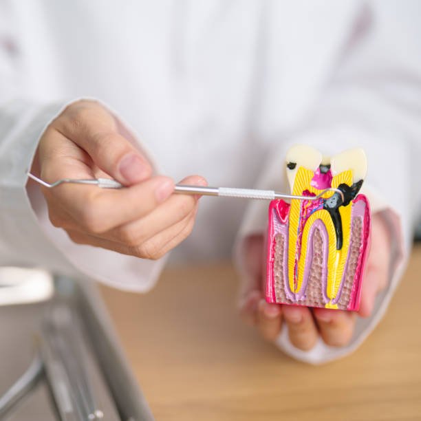
Dental Caries; also known as tooth decay or a cavity, is an infection, bacterial in origin, that causes demineralization and destruction of the hard tissues (enamel, dentin and cementum). Usually by production of acid by bacterial fermentation of the food debris accumulated on the tooth surface. If demineralization exceeds saliva and other remineralization factors such as from calcium and fluoridated toothpastes. These hard tissues progressively break down, producing dental caries (cavities, holes in the teeth). The bacteria most responsible for dental cavities are the mutans streptococci, most prominently Streptococcus mutans and Streptococcus sobrinus, and lactobacilli. If left untreated, the disease can lead to pain, tooth loss and infection. Today, caries remain one of the most common diseases throughout the world. The presentation of caries is highly variable. However the risk factors and stages of development are similar. Initially it may appear as a small chalky area (smooth surface caries), which may eventually develop into a large cavitation. Sometimes caries may be directly visible. However other methods of detection such as X-rays are used for less visible areas of teeth and to judge the extent of destruction. tooth restoration to minimize the chance of recurrence. Signs and symptoms for Dental Caries A person experiencing caries may not be aware of the disease. The earliest sign of a new carious lesion is the appearance of a chalky white spot on the surface of the tooth, indicating an area of demineralization of enamel. This is referred to as an incipient carious lesion .As the lesion continues to demineralize, it can turn brown but will eventually turn into a cavitation (“cavity”). Before the cavity forms the process is reversible, but once a cavity forms the lost tooth structure cannot be regenerated. A lesion that appears brown and shiny suggests dental caries was once present but the demineralization process has stopped, leaving a stain. A brown spot that is dull in appearance is probably a sign of active caries. As the enamel and dentin are destroyed, the cavity becomes more noticeable. The affected areas of the tooth change color and become soft to the touch. Once the decay passes through enamel, the dentinal tubules, which have passages to the nerve of the tooth, become exposed, resulting in a toothache. The pain may worsen with exposure to heat, cold, or sweet foods and drinks. Dental caries can also cause bad breath and foul tastes. Prevention Oral hygiene Personal hygiene care consists of proper brushing and flossing daily. The purpose of oral hygiene is to minimize any etiologic agents of disease in the mouth. The primary focus of brushing and flossing is to remove and prevent the formation of plaque. Plaque consists mostly of bacteria. As the amount of bacterial plaque increases, the tooth is more vulnerable to dental caries when carbohydrates in the food are left on teeth after every meal or snack. A toothbrush can be used to remove plaque on accessible surfaces, but not between teeth or inside pits and fissures on chewing surfaces. When used correctly, dental floss removes plaque from areas that could otherwise develop proximal caries. Other adjunct hygiene aids include interdental brushes, water picks, and mouthwashes. Professional hygiene care consists of regular dental examinations and cleanings. Sometimes, complete plaque removal is difficult, and a dentist or dental hygienist may be needed. Along with oral hygiene, radio-graphs may be taken at dental visits to detect possible dental caries development in high risk areas of the mouth. Dietary modification For dental health, frequency of sugar intake is more important than the amount of sugar consumed. In the presence of sugar and other carbohydrates, bacteria in the mouth produce acids that can demineralize enamel, dentin, and cementum. The more frequently teeth are exposed to this environment the more likely dental caries are to occur. Therefore, minimizing snacking is recommended, since snacking creates a continuous supply of nutrition for acid-creating bacteria in the mouth. Also, chewy and sticky foods (such as dried fruit or candy) tend to adhere to teeth longer, and, as a consequence, are best eaten as part of a meal. Brushing the teeth after meals is recommended. It has been found that milk and certain kinds of cheese like cheddar cheese can help counter tooth decay if eaten soon after the consumption of foods potentially harmful to teeth. Also, chewing gum containing xylitol (a sugar alcohol) is widely used to protect teeth in some countries. Other measures The use of dental sealants is a means of prevention. A sealant is a thin plastic-like coating applied to the chewing surfaces of the molars to prevent food from being trapped inside pits and fissures. This deprives resident plaque bacteria carbohydrate preventing the formation of pit and fissure caries. Sealants are usually applied on the teeth of children, shortly after the molars erupt. Sealants can wear out and fail to prevent access of food and plaque bacteria inside pits and fissures and need to be replaced. Calcium, as found in food such as milk and green vegetables, is often recommended to protect against dental caries. It has been demonstrated that calcium and fluoride supplements decrease the incidence of dental caries. Fluoride helps prevent decay of a tooth by binding to the hydroxyapatite crystals in enamel. The incorporated calcium makes enamel more resistant to demineralization and, thus, resistant to decay. Topical fluoride is also recommended to protect the surface of the teeth. This may include a fluoride toothpaste or mouthwash. Many dentists include application of topical fluoride solutions as part of routine visits. Treatment For Dental Caries For the small lesions, topical fluoride is sometimes used to encourage remineralization. For larger lesions, the progression of dental caries can be stopped by treatment. The goal of treatment is to preserve tooth structures and prevent further destruction of the tooth. Aggressive treatment, by filling, of incipient carious lesions, places where there is superficial damage to the enamel, is controversial as they may heal themselves, while once a filling is performed it will eventually have to be
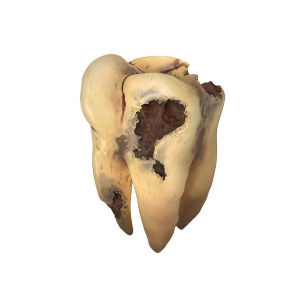
Tooth Wear: Abrasion, Attrition, Abfraction and Erosion. Tooth wear It is not uncommon for me to see excessive tooth wear on patients. It can happen with all ages but is more common in older patients. Tooth wear can be a serious dental problem but the good news is that for the most part it is preventable. The most common cause of tooth wear is abrasion. This is typically caused by using too much force while brushing your teeth. It can be complicated by using abrasive toothpaste especially. Those that promote teeth whitening. These toothpastes work by an abrasive action to remove extrinsic stains. While they can help to remove tea and coffee stains, they can also remove your tooth enamel. So someone that has a history of abrasion should not use whitening toothpaste, and consider brushing with a fluoride or xylitol mouth rinse INSTEAD of toothpaste. The next most common cause of tooth wear is attrition. Attrition is caused by grinding and clenching your teeth. Patients who grind their teeth at night typically cause the most damage. The affects of nightly grinding or bruxism can be greatly reduced by wearing a night guard while you sleep. A night guard is a generic term for an appliance placed on your upper or lower teeth to prevent tooth wear. Chemical erosion Chemical erosion is also common, especially in those patients that have a low ph in their saliva. Soft drinks probably being the most common factor. But many other contributing factors can cause a low ph and erosion as well. Such as: sucking on lemons or too much lemon in their water, acid reflux, bulimia and sugar to name a few. After one drinks a soda pop for example it is a good idea to rinse out their mouth with water to raise the ph. Even better is to rinse out with a xylitol rinse or chew a piece of xylitol gum which increases the salivary ph. Lastly there is the process of enamel wear called abfraction. This is caused by the flexing of the tooth during grinding. The thinnest area of enamel is at the root surface so this enamel progressively fractures off. Often times tooth wear is cause by a combination of these factors. It is important for your dentist to educate you early as to the causes. So that preventative measures can be taken. Sometimes restorative treatment is necessary but usually some lifestyle changes can make all the difference. European Dental Center; best dental clinic in Jordan provide this information about Tooth Wear.
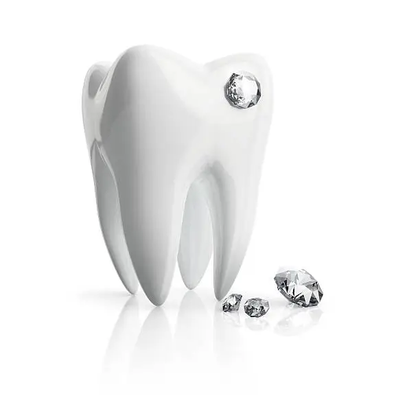
Tooth Jewelry is a simple way to add sparkle to your smile. Types of tooth Jewelry Common types of Tooth Jewelry: Tooth diamond. Tooth gold. Tooth tattoos. Tooth gems. Teeth decals. Tooth jewelry chain. Jewelry stabilizing Procedure & bonding instructions: The tooth is cleaned with a fluoride-free polishing paste. Completely dry and isolate the tooth. The dentist etches the tooth with 37% orthophosphoric acid for about 20-30 seconds to increase the surface area for bonding. Rinse surface thoroughly with water and blow dry for 10 sec. (no etchant should remain on the tooth!) Apply a light-curing bonding agent. Leave it on for a maximum of 20 seconds, distribute bonding through air blowing. Then light-cure for 20 sec. Apply a small amount of flow composite to the surface of the tooth. Use a jewel handler to easily pick up the jewel. Press it into the center of the composite. Ensure that the composite oozes on the sides, encircling the jewel with the composite to guarantee macro mechanical retention, while also ensuring that the jewel makes direct contact with the enamel. Take the light-curing lamp and start curing the composite from the top for about 60 seconds The total time for jewel to set into the composite is 20 sec. Do not touch the jewel with your fingers once it’s removed from the case. To guarantee maximum adhesiveness, it is essential to avoid skin contact with the special coating on the backside of the jewel It takes about 4 minutes to safely affix the jewel. The application of tooth jewelry should be performed by a trained dental professional to ensure proper placement and minimize the risk of damage to the tooth enamel. Additionally, individuals should maintain proper oral hygiene practices to prevent any complications such as tooth decay or gum irritation. European Dental Center; best dental clinic in Jordan provide this information about Tooth Jewelry.
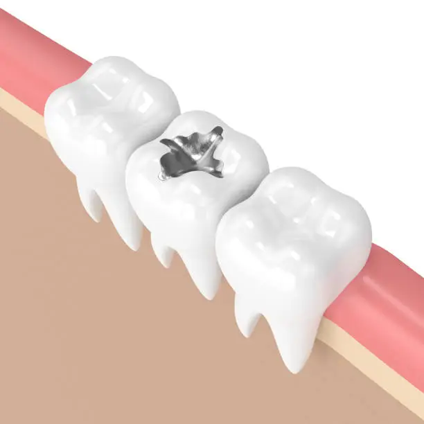
Should I replace my silver fillings with white fillings? Many patients want to replace their old silver fillings with white, natural-looking restorations. And many patients also ask, “Which one is better?” Should I replace my silver fillings with white fillings? This article seeks to educate patients about composites or “white fillings” and amalgam or “silver fillings”. Amalgams Dental amalgam, or “silver fillings”, has been used by dentists for the past 150 years. Dental amalgam consists of roughly a 50/50 mix of mercury and an alloy powder. Usually composed of silver, zinc or palladium. It has been the most tested dental material to date. Recently, its safety as a filling material has been questioned due to its mercury content. Many people believe that the mercury contained in the amalgam is toxic and could cause several health issues. Whether or not this amount of mercury is harmful to the body is a subject of controversy. Most people have some silver amalgam fillings in their teeth with no apparent adverse effects. No harm from the mercury in amalgam fillings has ever been absolutely proven. Additionally, once the silver filling is placed by the dentist, the patient has to wait 24 hours to eat on that side of the mouth, otherwise the filling could crumble and break. It is well known that amalgams do last for many, many years. However, after many years of the silver filling being in your mouth, they allow saliva under them and can corrode. Also, the tooth flexes around the amalgam during mastication, causing small cracks that with time can cause breakage within the tooth and finally it can fracture the tooth. Generally, after a large silver filling breaks, the tooth needs a crown in order to protect the tooth from breaking further. Amalgam fillings have a dark, gray appearance and many people do not like the look of their mouth when they smile or talk. Amalgams are a good option for cases where esthetics is not a concern and it is not possible to keep the area dry in order to bond a white filling. They are also generally less expensive than white fillings. Composites Composites or “white fillings” are formed from polymers, forming a hard plastic. The term “composite” means “made up of distinct parts or elements”. Therefore, a composite filling is basically a mix of polymers or plastic materials and fillers such as quartz, silica or barium. These fillers provide the strength to the composite material. Composite fillings come in a variety of colors. So they can be matched to the color of the patient’s own teeth. Composite fillings also lend strength to the tooth, whereas amalgams don’t. After the filling is light cured in the tooth, the patient can eat on it right away, since the material is completely set by the light, while the patient is in the dentist’s office. Also, composites require less removal of tooth structure because they are chemically bonded to the cavity preparation essentially splinting the tooth together. Because of this fact (chemical retention), we only remove the decayed portion of the tooth. This is in contrast to silver fillings, in which more healthy tooth often has to be removed in order for the amalgam to mechanically stay in place. Composite fillings have a natural, “white” appearance and many people prefer the look of their mouth when they smile or talk. Composites are an excellent option for cases where esthetics is a concern and it is possible to keep the area dry in order to bond a white filling. However, they are generally more expensive than amalgams. Are composite fillings weaker than amalgams? In short, yes, they are. The silver filling by itself is a stronger material, although it weakens the tooth. If you look at the total result — the filling plus the tooth —composites are stronger because they bond to the tooth. Making the tooth more resistant to fracture while old amalgams tend to break the tooth. I would rather have a tooth that lasts in the mouth. Rather than a filling that last in an extracted tooth. I’ve heard I should get my silver fillings replaced. When is this a good idea? We recommend replacement of silver fillings when the tooth is susceptible to fracture. When we suspect that there is a cavity under the existing filling. This can occur most commonly in old silver fillings. Silver fillings tend to hide cavities. Which means that you may have a cavity under a silver filling. And it will not show up on an x-ray. That is why we recommend that patients get periodic dental exams and x-rays. So we can detect cavities early and maintain your oral health. European Dental Center; best dental clinic in Jordan provide this information about Should I replace my silver fillings with white fillings?.


